01
«Асимметрия лица»
Асимметрия лица. Фиксируем во время диагностики и связываем с подходящим путём лечения.
 Записаться
Дермальный филлер может смещаться из исходной точки в окружающие ткани, меняя контур и создавая асимметрию - путь коррекции под контролем УЗИ.
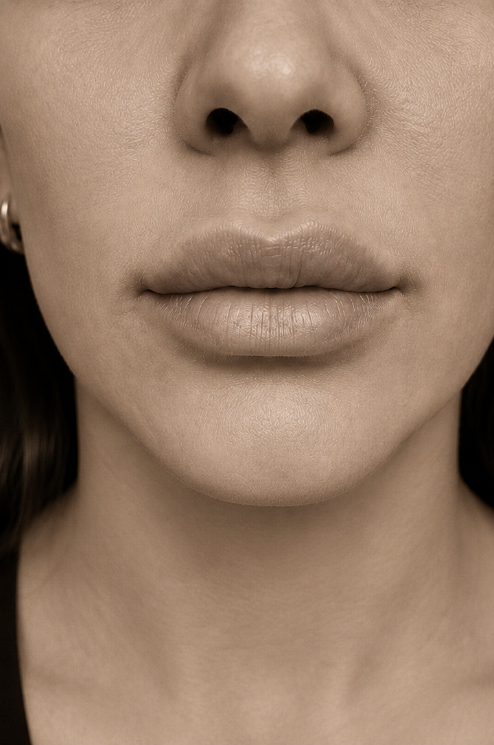 До · Изменение заметно
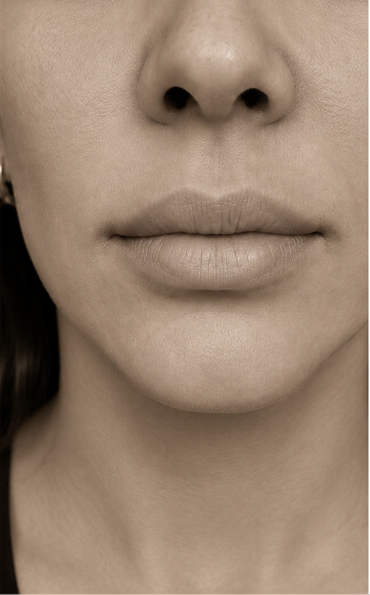 После · Восстановлено
Миграция филлера происходит, когда дермальный филлер смещается из первоначальной точки введения в окружающие ткани, изменяя контур и создавая асимметрию или неестественный вид.
Часто связано с избыточным объёмом филлера, неверной техникой введения, неподходящим выбором продукта или анатомическими особенностями. Коррекция выполняется аккуратно и под контролем визуализации.
"Нельзя исправить то, чего не видишь. УЗИ превращает миграцию филлера из догадки в план."
Формулировки меняются, но закономерность одна. Каждая из них - клинический сигнал, к которому мы прислушиваемся на консультации.
Асимметрия лица. Фиксируем во время диагностики и связываем с подходящим путём лечения.
Потеря чёткости губ или щёк. Фиксируем во время диагностики и связываем с подходящим путём лечения.
Прибавка объёма в непредназначенной зоне. Фиксируем во время диагностики и связываем с подходящим путём лечения.
Слегка изменилось по сравнению с прошлым годом. Фиксируем во время диагностики и связываем с подходящим путём лечения.
Фото ловит это чётче, чем зеркало. Фиксируем во время диагностики и связываем с подходящим путём лечения.
Ощущается иначе даже до того, как выглядит иначе. Фиксируем во время диагностики и связываем с подходящим путём лечения.
Редко бывает что-то одно. Путь строится вокруг наиболее активного фактора.
Миграция филлера становится очевидной только после визуализации. УЗИ мягких тканей - стартовая точка, план коррекции строится по тому, что показал скан.
Одна цель: работать с причиной, а не только с поверхностью. Основной протокол работает изнутри; поддерживающие пути углубляют или продлевают результат.
Инъекция фермента для растворения смещённого филлера на основе гиалуроновой кислоты - необходимый первый шаг перед любым повторным введением.
Когда мигрировавший продукт растворён, новый план использует правильную глубину, продукт и объём.
Поддерживающее · Качество кожиУвлажнение после коррекции, чтобы ткань восстановилась, а поверхность стала лучше.
Структурное · Долгосрочная плотностьТам, где миграция открывает структурную дряблость, стимуляторы восстанавливают опору.
Консультация · Полный сбросПлан под руководством врача, чтобы пересмотреть весь протокол лица, а не поправить одну зону.
Врач Beauté Concept осматривает кожу, определяет причину проблемы и разрабатывает протокол - от домашнего ухода до клинического курса.